
My name is Jaap, and I am a biomedical scientist that also used to live with severe Hidradenitis Suppurativa, the kind that takes over your life. Today, I am completely asymptomatic because I learned how to heal Hidradenitis Suppurativa from within. More importantly, I’ve had the privilege of helping many other individuals with HS get their lives back too.
A Proven natural Roadmap to Manage HS
Get the support and natural strategies you need for lasting relief and join a community that understands.

Introduction: The Hidden Architects of Your Skin
Have you ever felt like your body is a black box? Like the inflammation and pain of Hidradenitis Suppurativa (HS) comes out of nowhere, without rhyme or reason? It’s a frustrating, isolating feeling that I know all too well. For years, the conventional approach has focused on the symptoms, the lesions, the pain, the scarring. But what if the real story, the key to understanding how to treat Hidradenitis Suppurativa, lies deeper within the very architecture of our skin?
A groundbreaking new study just published in Nature Immunology has given us an unprecedented look into this hidden world (1). Using incredibly advanced technology, a team of researchers has created a detailed map of the skin’s fibroblasts—the cells responsible for building the skin’s framework, healing wounds, and, as it turns in, directing the immune system. They didn’t just look at healthy skin; they analyzed skin from 23 different diseases, including our own battleground: Hidradenitis Suppurativa.
What they found is a game-changer. It confirms a core philosophy we live by at HS Arma: HS isn’t just a skin disease. It’s a systemic issue, and to find a natural treatment of HS that truly works, we have to look at the root cause of the inflammation. This research gives us a powerful new lens through which to see our own healing journey. Let’s dive in and explore what these architect cells are telling us.
The Study: Mapping the Skin’s Cellular Neighborhood
First off, let’s give a huge thank you to the researchers behind this monumental effort. They integrated data from over 2.1 million individual cells to build this atlas. Think of it like creating a Google Maps for our skin, but instead of streets and buildings, we’re seeing individual cell types and where they live. It’s a stunning achievement that provides incredible clarity.
Before this, we knew fibroblasts existed, but we didn’t fully appreciate that there were so many different types, each with a specific job. The researchers identified six major fibroblast subtypes in healthy skin and, crucially, three more that are specific to diseased skin. Two of these are incredibly relevant to our fight with HS.
A Proven natural Roadmap to Manage HS
Get the support and natural strategies you need for lasting relief and join a community that understands.

Figure 1: A New Map of Our Skin’s Fibroblasts
Figure 1 Legend: An Overview of the Fibroblast Atlas. This figure is the master plan for the entire study. Figure by Steele and colleagues
Panel A shows the workflow: the researchers took 32 different datasets of human skin cells (totaling 2.1 million cells), used a computational method called “scVI integration” to combine them, and then focused on the 357,276 cells they identified as fibroblasts. They created a “healthy/nonlesional reference” map and then used it to analyze fibroblasts from 23 different skin diseases.
Panel B is a UMAP plot, a way of visualizing complex data. Each dot is a single fibroblast from healthy skin, and they are clustered together based on how similar their gene expression is. The different colors represent the distinct fibroblast subtypes the researchers identified, like the “F1: superficial” in yellow or the “F3: FRC-like” in pink.
Panel C is a dot plot showing the specific genes that act as markers for each subtype. The size of the dot shows what percentage of cells in that group express the gene, and the color intensity shows the average expression level. This is the genetic fingerprint for each cell type.
Panel D provides a simple, illustrated summary of the main fibroblast types and their proposed functions, such as the “F3: FRC-like” cells being involved in immune cell communication.
A Proven natural Roadmap to Manage HS
Get the support and natural strategies you need for lasting relief and join a community that understands.
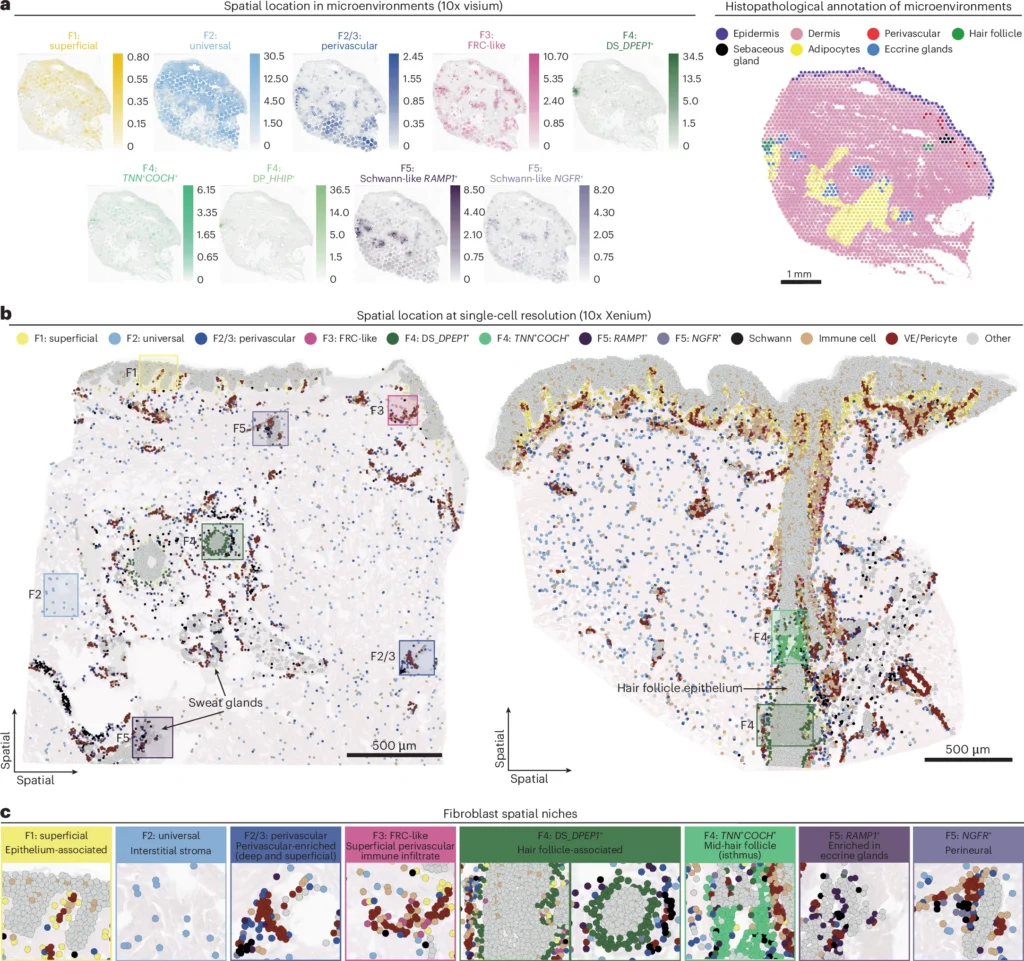
Figure 2: Finding Where Fibroblasts Live
Figure 2 Legend: Pinpointing Fibroblast Niches in the Skin. This figure shows the spatial location of these different fibroblast subtypes—where they actually live in the skin. This was done using two powerful technologies, 10x Visium and 10x Xenium, which measure gene activity directly in slices of tissue. Figure by Steele and colleagues
Panel A shows Visium data. The heatmaps on the left show the abundance of each fibroblast subtype’s gene signature across a skin sample. For example, you can see “F1: superficial” fibroblasts are concentrated right under the epidermis. The image on the right is a histopathological map of the same tissue, annotating structures like hair follicles and glands.
Panel B uses the higher-resolution Xenium technology to map individual cells. Each colored dot represents a single cell type, allowing us to see exactly which cells are neighbors. You can see clusters of F3 (pink) and F2/3 (blue) fibroblasts around blood vessels (perivascular regions).
Panel C zooms in on these “spatial niches,” showing how specific fibroblast subtypes are associated with structures like hair follicles (F4, green) or eccrine glands (F5, purple). This spatial context is crucial because it tells us who these fibroblasts are “talking” to and what their function might be.
The Two Faces of Fibroblasts in HS: Healers and Attackers
Here’s where it gets really interesting for us HS warriors. The study found that in diseased skin, these fibroblast populations change dramatically. They identified two “disease-adapted” subtypes and three “disease-specific” subtypes that barely exist in healthy skin. Two of these are central to the HS story.
1. F3: The “Immune-Organizing” Fibroblast (FRC-like)
In healthy skin, there’s a type of fibroblast called F3: FRC-like. These cells are amazing. They act like little command centers, organizing immune cells in the skin, much like specialized cells do in our lymph nodes. They release signals (like the chemokines CCL19 and CXCL12) that call immune cells to the right place and tell them what to do.
In inflammatory diseases like HS, these F3 cells go into overdrive. They become “activated,” sending out even stronger signals that recruit more and more immune cells to the area. This is a critical piece of the puzzle. It helps explain why HS lesions become so intensely inflamed—our own skin’s infrastructure is calling in the troops.
2. F6: The “Inflammatory-Attacker” Myofibroblast
This is the big one. The researchers discovered a disease-specific subtype they call F6: inflammatory myofibroblasts. These cells are almost completely absent in healthy skin but are found in high numbers in diseases with a risk of scarring and chronic inflammation, like HS, acne, and even skin cancer.
These F6 cells are trouble. They don’t just build tissue; they actively fuel the fire of inflammation. They churn out a cocktail of inflammatory signals (like CXCL5, CXCL8, and MMP1) that specifically recruit neutrophils and monocytes, two key drivers of the tissue destruction we see in HS tunnels and abscesses.
Think of it this way: The F6 fibroblast is like a construction worker who suddenly starts screaming for the demolition crew to come in and tear the place down. This is the “fire” in our analogy, and it’s happening right within the structural cells of our own skin.
A Proven natural Roadmap to Manage HS
Get the support and natural strategies you need for lasting relief and join a community that understands.

Figure 3: Identifying the Disease-Specific Fibroblasts
Figure 3 Legend: Discovering New Fibroblast States in Diseased Skin. This figure explains how the researchers found the new, disease-associated fibroblast types. Figure by Steele and colleagues
Panel A shows their “scPoli” machine learning method. They trained a model on healthy cells and then used it to label cells from diseased skin. The cells that didn’t fit any healthy profile were marked as “uncertain.”
Panel B shows the results. On the left, you see the initial predictions, with a large gray area of uncertain cells. On the right, after re-clustering these uncertain cells, new subtypes emerge, including the F6, F7, and F8 myofibroblasts.
Panel C is a dot plot showing the unique gene markers for these disease-specific cells, such as the inflammatory chemokines (CXCL5, IL11) in F6 cells and matrix-remodeling genes (COL8A1, SFRP4) in F7/F8 cells.
Panel D graphically shows the density of fibroblasts, highlighting how the myofibroblast populations (F6/7/8) appear in diseased skin where they were absent in healthy skin.
Panels E and F summarize the changes, showing how healthy F1 and F3 cells become “activated” in disease, and detailing the new disease-specific types like the F6 inflammatory myofibroblast.
Panels G and H provide more detail on the gene signatures, showing the specific inflammatory and tissue-remodeling genes that these myofibroblasts turn on.
What This Means for You: The HS Arma Philosophy in Action
So, how does this cutting-edge science help us answer the question, can you cure HS?
This is where we see the profound gap between conventional medicine and a root-cause approach. The current medical model sees the inflammation, the neutrophils, the cytokines—and tries to block it with drugs. It’s like hearing a smoke alarm and just cutting its wires. You’ve stopped the noise, but the fire is still burning. Pharmaceuticals can be a powerful and necessary temporary shield, but they don’t put out the fire.
The HS Arma philosophy is different. We believe the focus should be on stopping the fire itself. This research shows us that the fire is being fueled by these F6 inflammatory myofibroblasts. The critical question then becomes: What is causing these fibroblasts to turn rogue in the first place?
The paper doesn’t answer this directly, but it points us exactly where we’ve been looking all along: systemic, body-wide inflammation. These fibroblasts are responding to danger signals in their environment. Those signals are the metabolic dysfunction, the gut dysbiosis, the nutrient deficiencies, and the chronic stress that we know are the upstream drivers of HS.
This is why at HS Arma, we focus on highly effective evidence-based nutrition and lifestyle change, and natural therapies and practices. We are building our defenses, layer by layer, to remove the triggers that cause these fibroblasts to become inflammatory.
- Foundational Nutrition: By removing inflammatory foods and embracing an anti-inflammatory diet, we stop sending the danger signals that activate these F6 cells.
- Strategic Lifestyle Changes: By managing stress and improving sleep, we calm the systemic alarm bells that put our immune system, and our fibroblasts, on high alert.
- Natural Therapies: By using targeted, science-backed natural compounds, we can directly reduce systemic inflammation and support the body’s return to balance.
This research powerfully validates our approach. The goal isn’t just to block a single inflammatory pathway; it’s to create an internal environment of health where our fibroblasts never get the signal to become inflammatory attackers in the first place. This is how we achieve deep, lasting remission.
A Proven natural Roadmap to Manage HS
Get the support and natural strategies you need for lasting relief and join a community that understands.
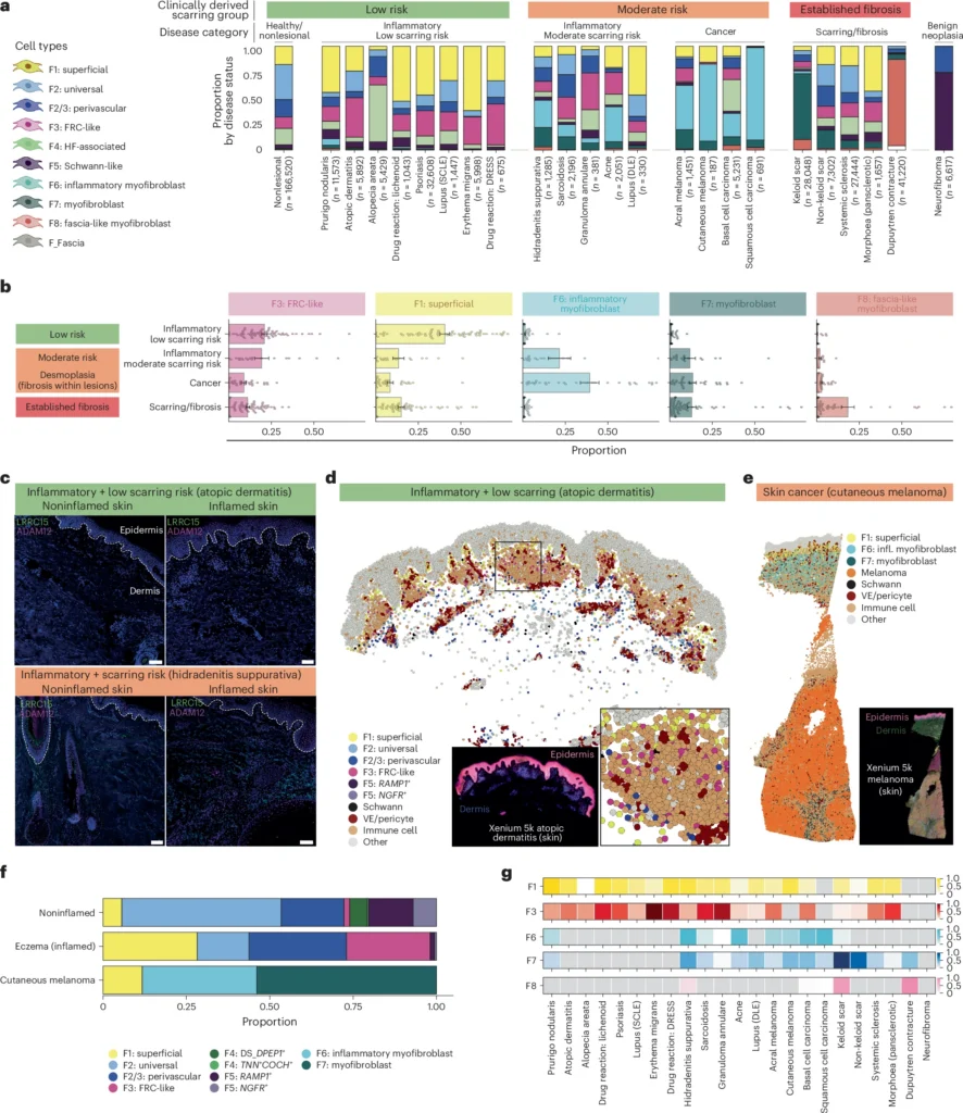
Figure 4: Linking Fibroblast Types to Scarring Risk
Figure 4 Legend: Fibroblast Composition Predicts Disease Outcome. This figure powerfully connects fibroblast subtypes to clinical outcomes. Figure by Steele and colleagues
Panel A categorizes 23 skin diseases into groups based on their risk of scarring. You can see Hidradenitis Suppurativa is in the “Moderate risk” inflammatory category. The stacked bars show the proportion of each fibroblast subtype in each disease.
Panel B averages these proportions and shows a striking pattern: diseases with low scarring risk are dominated by activated F1 and F3 cells, while diseases with moderate scarring risk (like HS) and cancer have a high proportion of F6 inflammatory myofibroblasts. Established scars are dominated by F7 myofibroblasts.
Panel C provides visual proof using immunofluorescence. The green stain (LRRC15) marks myofibroblasts. You can see it’s brightly lit up in inflamed HS skin but absent in non-inflamed skin and even in inflamed atopic dermatitis (a low-scarring-risk disease).
Panels D and E use spatial transcriptomics to visualize the stroma in low-risk vs. moderate-risk disease, showing the F6/F7 myofibroblast takeover in skin cancer.
Panel F confirms the proportional differences seen in the larger dataset.
Panel G shows that the gene signature for F6 cells is highest in diseases like HS, acne, and skin cancers, reinforcing their link to inflammatory, scarring-prone conditions.
The Life Cycle of a Scar: From Inflammation to Fibrosis
The study also gives us incredible insight into how HS tunnels and scars form. By looking at wound healing over 30 days, they saw a clear progression:
- Day 1-7: The F6 inflammatory myofibroblasts appear first and quickly dominate the scene, calling in the demolition crew (neutrophils, monocytes).
- Day 30: The F6 cells fade, and another type, F7 myofibroblasts, takes over. These F7 cells are less inflammatory and more focused on producing massive amounts of collagen and creating the dense, hard tissue of an established scar.
This suggests that the F6 cells are an intermediate state. They create the inflammatory chaos, and then they transition into F7 cells to lay down the scar tissue. This is the tragic, two-act play of an HS lesion. By stopping the F6 cells from ever taking the stage, we can potentially prevent the scarring that follows.
A Proven natural Roadmap to Manage HS
Get the support and natural strategies you need for lasting relief and join a community that understands.

Figure 5: The Journey to a Myofibroblast
Figure 5 Legend: Tracing the Origins of Scar-Forming Cells. This figure uses computational methods to predict how healthy fibroblasts turn into disease-specific myofibroblasts. Figure by Steele and colleagues
Panel A, B, and C use different algorithms (“velocity pseudotime,” “PAGA,” and “CellRank”) to map the likely differentiation paths. The arrows consistently show two main routes: one where F2 “universal” fibroblasts can become F7 myofibroblasts directly, and another where F1 “superficial” fibroblasts first turn into F6 “inflammatory” myofibroblasts, which then mature into F7s. This highlights F6 as a key intermediate state.
Panel D validates this prediction using real data from a human wound healing study. In baseline (unwounded) skin, there are no F6 or F7 cells. But by Day 7 post-wounding, F6 inflammatory cells are the dominant type. By Day 30, the F7 scar-forming myofibroblasts have taken over. This is a real-time snapshot of the trajectory predicted by the models.
Panel E provides a schematic summary of this predicted journey from healthy skin to a wounded/inflamed state, culminating in the formation of scar tissue.
The Ultimate Proof: Why HS is a Whole-Body Problem
Now, for the part that I find most validating for our community. The researchers asked a critical question: “Are these rogue F6 inflammatory fibroblasts only a skin problem?” The answer is a resounding “NO,” and it’s the ultimate scientific proof for why we must treat HS from the inside out.
The researchers expanded their search and looked for these same fibroblast subtypes in other diseases affecting completely different parts of the body, like the gut in Inflammatory Bowel Disease (IBD) or the joints in Rheumatoid Arthritis. And what did they find? The exact same F6 inflammatory myofibroblasts, with the same genetic signature, fueling the fire of inflammation in those tissues, too.
This is the smoking gun. It proves that the cellular chaos driving our HS lesions is not some freak, isolated event in the skin. It is a fundamental pattern of systemic inflammation. The same internal environment that can trigger these rogue fibroblasts in the gut of an IBD patient can trigger them in the skin of an HS warrior.
A Proven natural Roadmap to Manage HS
Get the support and natural strategies you need for lasting relief and join a community that understands.
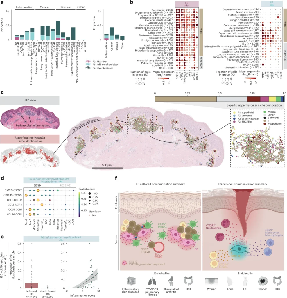
Figure 6: A Shared Blueprint for Inflammation Across the Body
Figure 6 Legend: A Shared Pattern of Inflammation Across the Body. This figure is the ultimate proof that HS is a systemic disease. The researchers looked for the disease-associated fibroblasts in other tissues beyond the skin. Figure by Steele and colleagues
Panels A and B show that F6 inflammatory myofibroblasts are not just a skin problem; they are abundant in other inflammatory conditions like IBD, lung disease, and cancer. The dot plot for F6 cells in
Panel B is particularly telling: it shows that the genetic signature for these inflammatory cells in Hidradenitis Suppurativa is remarkably similar to the one found in IBD and early wounds. Panel D and the schematic in
Panel F (right side) summarize the key function of these F6 cells: they release a specific cocktail of signals (like CXCL5 and CXCL13) to call in neutrophils and monocytes, the very immune cells that cause the destructive inflammation in HS lesions.
Panel E provides stunning clinical proof from IBD patients, showing that the number of these F6 fibroblasts in the gut tissue directly correlates with the severity of inflammation. More F6 cells means a worse flare-up. The message is clear: the F6 inflammatory myofibroblast is a key player in chronic, systemic inflammation, no matter which organ it affects.
This is why a foundational, natural approach is non-negotiable for lasting remission. You cannot hope to solve the problem by only focusing on the skin in your armpits or groin when the root signals that create these inflammatory cells are body-wide. You have to put out the systemic fire.
A Proven natural Roadmap to Manage HS
Get the support and natural strategies you need for lasting relief and join a community that understands.

Key Takeaways
For those of you skimming (I know you’re busy!), here are the most critical points:
- HS is not just skin deep: It involves the very cells that build and maintain your skin’s structure (fibroblasts).
- A “rogue” fibroblast exists: A specific subtype called the F6 inflammatory myofibroblast appears in HS. It actively recruits the immune cells that cause tissue damage and abscesses.
- This is a whole-body problem: The same rogue F6 fibroblasts are found in other systemic inflammatory diseases like IBD, proving that the root cause is not isolated to the skin.
- Inflammation precedes scarring: The F6 inflammatory cell appears first, creating chaos. It then likely turns into an F7 cell, which creates the final scar. Stopping the F6 cell is key to preventing scarring.
- You can control the signals: The appearance of these rogue F6 cells is a response to systemic inflammation. A root-cause approach focusing on diet and lifestyle can remove the triggers that create them.
Conclusion: You Are the Healer
This research is more than just a fascinating look at cell biology. It is a message of profound hope and empowerment. It tells us that the progression of HS isn’t some mysterious, unstoppable force. It is a biological process we can understand and, more importantly, that we can influence.
The path to healing isn’t about finding a single magic bullet to kill off one cell type. It’s about changing the entire environment. It’s about building your HS Armor, piece by piece, until your body is so resilient and balanced that your fibroblasts never get the call to go to war. This is a core principle we put into practice every day in our HS Arma model approach. Remission is possible, and the power to achieve it is already within you.
References
- Steele, L., Olabi, B., Roberts, K., Mazin, P. V., Koplev, S., Tudor, C., … & Haniffa, M. (2025). A single-cell and spatial genomics atlas of human skin fibroblasts reveals shared disease-related fibroblast subtypes across tissues. Nature Immunology. Published online 24 September 2025. https://doi.org/10.1038/s41590-025-02267-8
A Proven natural Roadmap to Manage HS
Get the support and natural strategies you need for lasting relief and join a community that understands.

Important Medical Disclaimer
1. Not Medical Advice: All content and information on this website is for informational and educational purposes only. It does not constitute medical advice and is not a substitute for professional diagnosis, treatment, or consultation with a qualified healthcare provider.
2. My Role and Qualifications: I am a biomedical scientist and PhD candidate and share information from that perspective, combined with my personal experience as a patient with Hidradenitis Suppurativa. However, I am not a medical doctor, physician, or registered healthcare professional. Do not consider our relationship a doctor-patient relationship.
3. Consult Your Doctor: Always seek the advice of your medical doctor or another qualified health professional with any questions you may have regarding a medical condition. Never disregard professional medical advice or delay seeking it because of something you have read on this website. If you suspect you are experiencing a medical emergency, or a severe infection, do not rely on this website or the HS Arma community, please call your local emergency services or go to the nearest emergency room immediately.
4. A Critical Warning on Medication: Pharmaceutical drugs are a crucial tool in managing Hidradenitis Suppurativa for many people. Under absolutely no circumstances should you ever alter, reduce, or stop taking your prescribed medication without the explicit direction of the doctor who prescribed it. Doing so can be dangerous. Always consult with your doctor before doing anything related to your treatment plan.
5. No Liability: Your use of this website and reliance on any information provided is solely at your own risk.
6. Individual Results May Vary: Every patient’s biological baseline, genetics, and adherence to the protocol is different. Therefore, I cannot guarantee specific results, cures, or timelines for your Hidradenitis Suppurativa.
7. Scientific and Expressive Freedom: The articles published on this blog are distinct from formal peer-reviewed academic literature. They serve as an independent platform for my personal viewpoints, scientific hypotheses, and philosophical reflections as an independent scientist and HS patient. While grounded in biomedical research, I exercise a degree of expressive freedom to translate rigid academic data into insights from a patient perspective. These writings are my personal meditations on the science of HS and should be read as my individual perspective, not as universally accepted clinical consensus or formal peer-reviewed literature.