My name is Jaap, and I am a biomedical scientist that also used to live with severe Hidradenitis Suppurativa, the kind that takes over your life. Today, I am completely asymptomatic because I learned how to heal Hidradenitis Suppurativa from within. More importantly, I’ve had the privilege of helping many other individuals with HS get their lives back too.
Are Overactive “Immune Guardians” Fueling Your Hidradenitis Suppurativa?
Today, I want to talk about a hidden player in the HS story. We often hear about blocked hair follicles and an overactive immune system, but what if I told you that a specific type of immune cell which is often not recognised, is being found right at the scene of the crime, fanning the flames of our most painful symptoms?
A fascinating new scientific paper has shed light on this, and it’s a perfect example of how we can use deep science to understand how to treat hidradenitis suppurativa from the inside out. This isn’t just theory; it’s a clue that points directly back to the foundational, natural approach we take to find lasting remission. Let’s break down what these researchers found and, more importantly, what it means for your healing journey.
Meet the Mast Cell: An Overlooked Player in the HS Drama
Before we dive into the study, let’s meet our main character: the mast cell.
Imagine the mast cell as a first responder or a security guard for your body’s tissues. It’s part of your innate immune system, embedded in your skin, waiting for trouble. When it detects a threat, like an injury or a pathogen, it sounds the alarm by releasing a flood of powerful chemicals, including histamine, tryptase, and chymase. This chemical alarm summons other immune cells to the area, creating inflammation to fight off the invader and begin the healing process.
In a healthy system, this is a beautiful, efficient process. But in autoimmune and chronic inflammatory conditions like HS, it goes haywire. The security guards become jumpy, paranoid, and trigger-happy. They start sounding the alarm constantly, even when there’s no real threat.
This is precisely what a brilliant team of researchers led by Dr. A. Flora and Dr. J. W. Frew investigated in their 2023 paper published in Experimental Dermatology. And I want to pause and thank them for their work. This is the kind of detailed, high-resolution science that helps us connect the dots.
A Proven natural Roadmap to Manage HS
Get the support and natural strategies you need for lasting relief and join a community that understands.
The research team wanted to know: what role are mast cells really playing in Hidradenitis Suppurativa tissue? They took skin biopsies from HS patients, from active lesions, the skin around tunnels (perilesional), and unaffected skin, and compared them. Here’s what they discovered.
Finding 1: The Alarms Are Blaring, HS Skin is Full of Activated Mast Cells
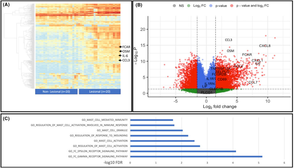
First, they looked at the overall genetic activity in the skin. Using a technique called RNA sequencing, they found that a whole host of genes and pathways related to mast cell activation were cranked up to high volume in HS lesions compared to unaffected skin.
Figure 1 shows this loud genetic signal. It confirms that the cellular machinery for mast cell activation is switched ON.
A Proven natural Roadmap to Manage HS
Get the support and natural strategies you need for lasting relief and join a community that understands.
Figure 1 Legend: Understanding the Genetic Blueprint of HS Inflammation. This figure gives us a bird’s-eye view of the genetic activity in HS skin. Figures from Dr. A. Flora and Dr. J. W. Frew.
(A) Heatmap: This colorful chart compares gene activity between non-lesional (left) and lesional (right) skin from 20 HS patients. Each row is a different gene, and each column is a patient sample. Red means a gene is highly active (“turned on”), while blue means it’s less active. You can see a dramatic shift to red in the lesional column, showing a massive inflammatory response. The arrows point to specific genes like FCAR, OSM, IL-6, and CCL3, which are all associated with mast cell and other innate immune cell activity.
(B) Volcano Plot: This is a statistical plot showing which genes are the most significantly changed. Genes on the right side are significantly upregulated (more active) in HS lesions. Many of the top hits are inflammatory messengers, confirming the intense immune reaction.
(C) Pathway Analysis: This bar chart shows which biological “pathways” or systems are most affected. Notice how many are related to mast cells: “MAST_CELL_MEDIATED_IMMUNITY,” “REGULATION_OF_MAST_CELL_ACTIVATION,” and “MAST_CELL_GRANULE.” This is powerful evidence that mast cell activity is a central part of the HS disease process. Figures from Dr. A. Flora and Dr. J. W. Frew.
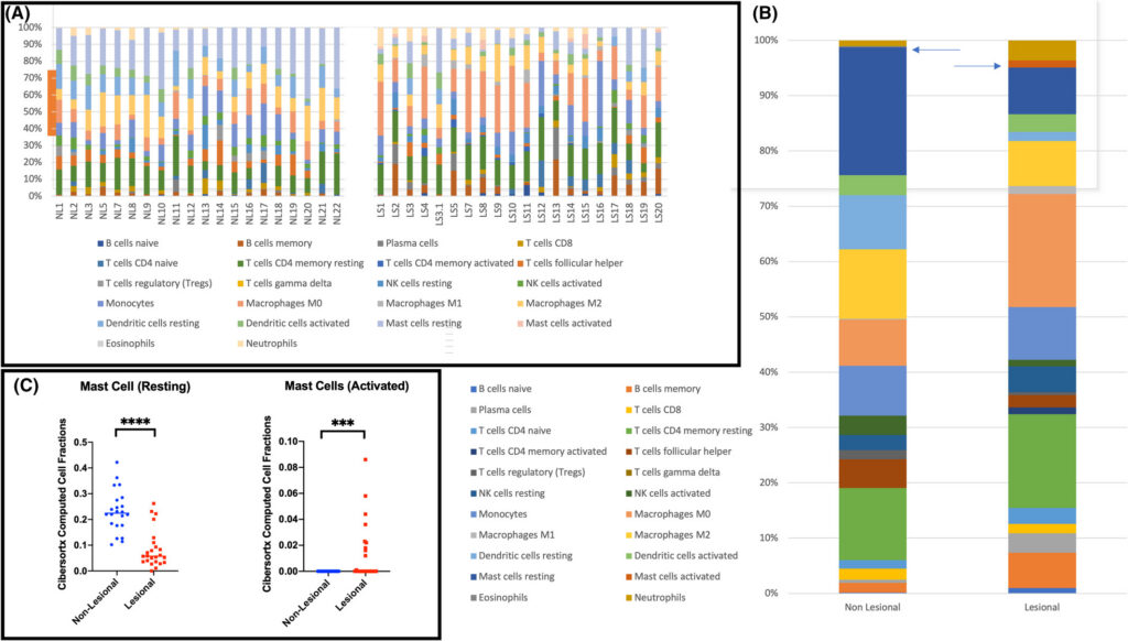
But they went deeper. Using a clever computational method, they estimated the different types of immune cells present. What they found was stunning. It wasn’t just that there were more mast cells overall; it was that the type of mast cell had changed dramatically. The proportion of “resting” mast cells plummeted, while the proportion of activated mast cells skyrocketed in lesional skin.
Figure 2 illustrates this perfectly. It’s not about the number of security guards on patrol; it’s about how many are actively blasting their sirens.
A Proven natural Roadmap to Manage HS
Get the support and natural strategies you need for lasting relief and join a community that understands.
Figure 2 Legend: Shifting from Resting to Activated Immune Cells. This figure uses a computational technique called CIBERSORTx to analyze the gene expression data from Figure 1 and estimate the proportions of different immune cell types. It’s like taking a census of the immune army in the skin. Figures from Dr. A. Flora and Dr. J. W. Frew.
(A) Individual Patient Data: These stacked bar charts show the immune cell makeup for each individual patient’s non-lesional (NL) and lesional (LS) skin samples. You can see the huge variability between people but also a clear trend: the lesional samples have much larger sections of B cells, plasma cells, and macrophages.
(B) Average Immune Profile: This chart averages the data from all patients. The Lesional bar on the right shows a dramatic increase in inflammatory cells compared to the Non-Lesional bar.
(C) The Key Finding for Mast Cells: This is the most important part. The graph on the left shows the fraction of Resting Mast Cells is significantly lower in lesional skin. The graph on the right shows the fraction of Activated Mast Cells is significantly higher. The takeaway is clear: in HS lesions, the mast cells are not just present; they are highly triggered and actively participating in the inflammatory cascade.
Finding 2: Location, Location, Location – Mast Cells Are Found at the Scene of the Crime
This is where it gets really interesting. The researchers used special stains to visualize exactly where these activated mast cells were hanging out in the skin.
They found them clustered in the most infamous HS locations:
Around the deep, painful tunnels that are so characteristic of the disease.
Alongside epithelial buds, which are the beginnings of new tissue growth and tunnel formation.
In fibrotic (scarred) regions deep in the dermis.
A Proven natural Roadmap to Manage HS
Get the support and natural strategies you need for lasting relief and join a community that understands.
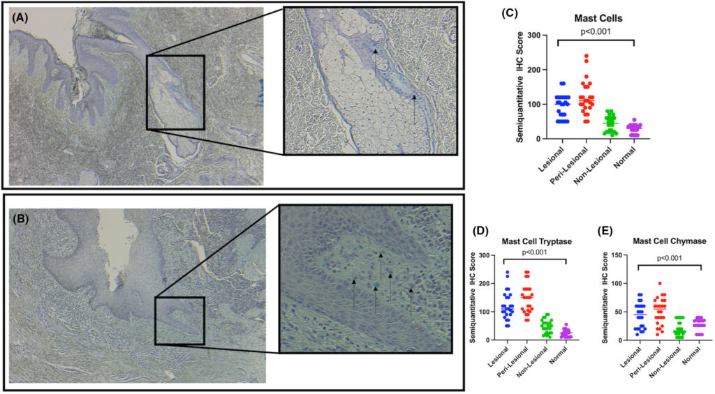
Figure 3 shows this visually. The mast cells aren’t just randomly scattered; they seem to be strategically positioned to contribute to tissue destruction, improper wound healing, and scarring.
Figure 3 Legend: Visualizing the Mast Cell Invasion. This figure uses a staining technique called immunohistochemistry (IHC), which uses antibodies to “light up” specific cells in a tissue sample, making them visible under a microscope. Here, they are staining for all mast cells. Figures from Dr. A. Flora and Dr. J. W. Frew.
(A) & (B) Mast Cells in Action: These microscopic images show mast cells (the small dark dots indicated by arrows) congregating around hair follicles and, more importantly, in and around the lining of an HS tunnel. The zoomed-in boxes give you a closer look. This shows they are physically present where the damage is happening.
(C), (D), & (E) Quantifying the Problem: These graphs count the stained cells. They show a statistically significant increase in the number of Mast Cells (C), as well as cells positive for their inflammatory contents Mast Cell Tryptase (D) and Mast Cell Chymase (E), as you move from normal skin to non-lesional, perilesional, and finally, full-blown lesional skin. This confirms that more mast cells are present and they are actively releasing their inflammatory cargo.
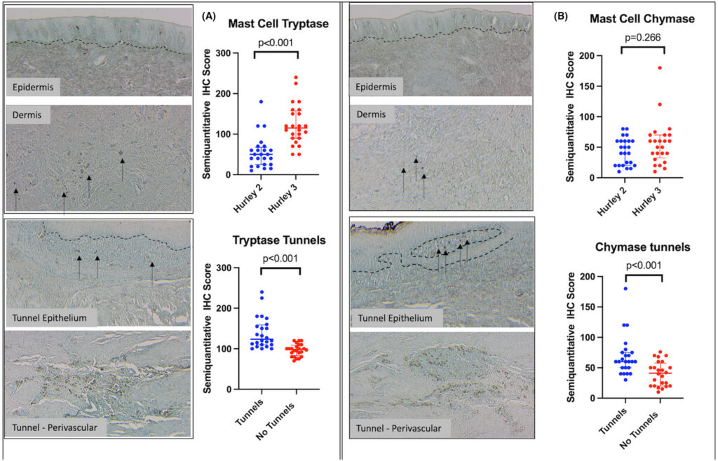
Furthermore, they found that the number of mast cells releasing their inflammatory contents was significantly higher in people with more severe (Hurley Stage 3) disease and in those with tunnels. This provides a direct link between mast cell activity and the clinical severity of HS.
A Proven natural Roadmap to Manage HS
Get the support and natural strategies you need for lasting relief and join a community that understands.
Figure 4 drives this point home, showing that the markers for mast cell degranulation (tryptase and chymase) are elevated right where the tunnels are.
Figure 4 Legend: Linking Mast Cells to Disease Severity and Tunnels. This figure dives deeper, staining for Tryptase and Chymase, two major enzymes released by mast cells when they degranulate or sound the alarm. Figures from Dr. A. Flora and Dr. J. W. Frew.
(A) Mast Cell Tryptase: The top left panel shows significantly more tryptase-positive mast cells in the skin of Hurley Stage 3 patients compared to Hurley Stage 2. The bottom left shows a increase in tryptase around tunnels compared to skin without tunnels. The images show these cells (dark dots) in the dermis and surrounding the tunnel epithelium.
(B) Mast Cell Chymase: The right panels show a similar story for chymase. While the difference between Hurley stages wasn’t statistically significant, the presence of chymase was higher in biopsies that contained tunnels.
The Big Picture: This tells us that not only are there more mast cells, but they are actively releasing their contents in the most damaged areas of HS skin, suggesting they play a direct role in the progression to more severe disease and the formation of tunnels.
Finding 3: A Drug Can Silence the Alarm… But Doesn’t Put Out the Fire
“The doctor of the future will give no medicine, but will instruct his patient in the care of the human frame, in diet and in the cause and prevention of disease.“ – Thomas Edison, 1903
Lastly, the researchers looked at what happened when patients were treated with a drug called Fostamatinib. This is a small molecule that inhibits an enzyme called Spleen Tyrosine Kinase (SYK), a crucial signaling molecule inside mast cells (and B cells). Think of it as cutting the power cord to the alarm system.
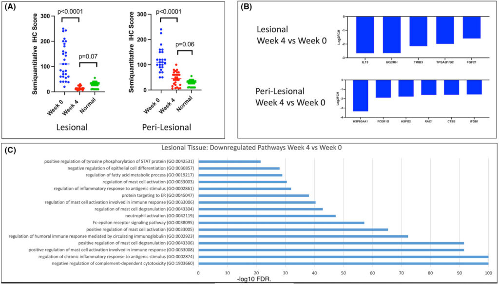
After just four weeks of treatment, they saw a dramatic reduction in the number of mast cells in lesional tissue, bringing them down to levels seen in normal, healthy skin. The genes and pathways associated with mast cell activation were also significantly quieted down.
A Proven natural Roadmap to Manage HS
Get the support and natural strategies you need for lasting relief and join a community that understands.
Figure 5 shows the powerful effect of this drug. This is an exciting finding for developing new treatments, and it proves that targeting mast cells can have a real impact.
Figure 5 Legend: The Effect of a Pharmaceutical “Shield.” This figure shows the results from a clinical trial where HS patients were treated with Fostamatinib, a drug that inhibits a key pathway (SYK) in mast cells. Figures from Dr. A. Flora and Dr. J. W. Frew.
(A) Reduction in Mast Cells: These graphs show the mast cell count in lesional and perilesional tissue at the start of the trial (Week 0) and after four weeks of treatment (Week 4). The number of mast cells drops dramatically, becoming statistically similar to the levels in normal, healthy skin. This shows the drug effectively removes these cells from the inflamed tissue.
(B) Quieting the Genetic Noise: These bar charts show the change in gene expression after 4 weeks. Genes associated with mast cell activation and inflammation (like IL13, TPSAB1/2 which codes for tryptase, and FCER1G) are significantly downregulated (turned off).
(C) Shutting Down Pathways: This chart shows that entire biological pathways related to mast cell activation, degranulation, and immune signaling are strongly suppressed by the treatment.
The Interpretation: This demonstrates that a pharmaceutical approach targeting this pathway is effective at silencing this specific aspect of HS inflammation. It’s a powerful temporary shield.
The Critical Question: Why Are the Alarms Blaring in the First Place?
This is where we bridge the gap between brilliant lab research and our real-world healing. The study is a masterpiece of “downstream” science. It identifies a problem (overactive mast cells) and shows that a drug blocking that problem works.
But this is like discovering that silencing a smoke alarm stops the annoying noise. It’s true, and it provides relief. But it never asks the most important question: what started the fire?
Why are the mast cells in our skin so chronically over-activated? Why are these security guards so trigger-happy?
The answer is that they are responding to a real, body-wide state of emergency: systemic inflammation. The source isn’t in the skin itself. The fire is upstream, caused by triggers from our diet, our gut health, chronic stress, poor sleep, and environmental exposures. This is the core philosophy of Autoimmune Arma. Conventional medicine focuses on managing the smoke, while we are dedicated to putting out the fire.
“The symptom is not the enemy, but the messenger.” – Dr. Lissa Rankin, 2013
A Proven natural Roadmap to Manage HS
Get the support and natural strategies you need for lasting relief and join a community that understands.
The hS Arma Approach: Treating HS by Extinguishing the Fire
This research doesn’t contradict our philosophy; it beautifully confirms it. It gives us a specific cellular mechanism that explains how the systemic inflammation we talk about creates the painful lesions we experience.
The Conventional View: See drugs and surgery as the main treatment. Lifestyle is a small, optional add-on. Their goal is to find a better way to silence the alarm (the SYK inhibitor).
The HS Arma View: We reverse this. We see that the natural approach is the main treatment because it addresses the root cause. At HS Arma, we focus on highly effective evidence-based nutrition and lifestyle change, and natural therapies and practices. The goal is to remove the inflammatory triggers so the mast cells (and the rest of the immune system) have no reason to sound the alarm in the first place.
Pharmaceuticals are powerful and sometimes necessary tools, a temporary shield to protect you while you do the foundational work. But our goal is to help you build such a strong foundation that naturally your cells behave the way they should and that these pharmaceutical shields become less necessary, or ideally, completely unnecessary. Seeing how these foundational changes have helped so many in our community truly brings this research to life.
Key Takeaways
A New Villain: Groundbreaking research has identified activated mast cells as key players in HS, found in high numbers around painful tunnels and scar tissue.
Fueling the Fire: These overactive immune security guards release inflammatory chemicals that contribute directly to the chronic inflammation and tissue damage of hidradenitis suppurativa.
Silencing the Alarm vs. Putting Out the Fire: While new drugs can block mast cells (silencing the alarm), this doesn’t address the root cause of why they are overactive.
The True Path to Healing: A lasting, natural treatment of HS must focus on identifying and removing the “upstream” triggers of systemic inflammation (the fire) through a holistic approach to diet, lifestyle, and targeted natural therapies.
This science is empowering. It gives us a deeper why behind the strategies we use to heal. It validates the pursuit of a root-cause solution. Healing is not about finding the perfect drug to silence a symptom; it’s about restoring balance to the entire body so that the symptoms have no reason to exist. Remission is possible.
“The human body is the most complex system ever created. The more we learn about it, the more we realize that the healing power of the body is more powerful than any drug ever invented.“ – Dr. Mark Hyman, 2014
A Proven natural Roadmap to Manage HS
Get the support and natural strategies you need for lasting relief and join a community that understands.
1. Not Medical Advice: All content and information on this website is for informational and educational purposes only. It does not constitute medical advice and is not a substitute for professional diagnosis, treatment, or consultation with a qualified healthcare provider.
2. My Role and Qualifications: I am a biomedical scientist and PhD candidate and share information from that perspective, combined with my personal experience as a patient with Hidradenitis Suppurativa. However, I am not a medical doctor, physician, or registered healthcare professional. Do not consider our relationship a doctor-patient relationship.
3. Consult Your Doctor: Always seek the advice of your medical doctor or another qualified health professional with any questions you may have regarding a medical condition. Never disregard professional medical advice or delay seeking it because of something you have read on this website. If you suspect you are experiencing a medical emergency, or a severe infection, do not rely on this website or the HS Arma community, please call your local emergency services or go to the nearest emergency room immediately.
4. A Critical Warning on Medication: Pharmaceutical drugs are a crucial tool in managing Hidradenitis Suppurativa for many people. Under absolutely no circumstances should you ever alter, reduce, or stop taking your prescribed medication without the explicit direction of the doctor who prescribed it. Doing so can be dangerous. Always consult with your doctor before doing anything related to your treatment plan.
5. No Liability: Your use of this website and reliance on any information provided is solely at your own risk.
6. Individual Results May Vary: Every patient’s biological baseline, genetics, and adherence to the protocol is different. Therefore, I cannot guarantee specific results, cures, or timelines for your Hidradenitis Suppurativa.
7. Scientific and Expressive Freedom: The articles published on this blog are distinct from formal peer-reviewed academic literature. They serve as an independent platform for my personal viewpoints, scientific hypotheses, and philosophical reflections as an independent scientist and HS patient. While grounded in biomedical research, I exercise a degree of expressive freedom to translate rigid academic data into insights from a patient perspective. These writings are my personal meditations on the science of HS and should be read as my individual perspective, not as universally accepted clinical consensus or formal peer-reviewed literature.