
My name is Jaap, and I am a biomedical scientist that also used to live with severe Hidradenitis Suppurativa, the kind that takes over your life. Today, I am completely asymptomatic because I learned how to heal Hidradenitis Suppurativa from within. More importantly, I’ve had the privilege of helping many other individuals with HS get their lives back, too.
Introduction: Listening to Your Skin’s Deepest Signals
Have you ever felt like your body is sending out distress signals, but no one seems to understand the message? If you’re living with Hidradenitis Suppurativa (HS), that feeling can be a constant companion. The painful nodules, the draining tunnels, the sheer exhaustion, it’s more than just a skin condition. It feels like something much deeper is fundamentally out of balance. We’re often handed antibiotics or offered biologics, treatments that try to silence the symptoms, but the root cause, the real reason why this is happening, often remains a frustrating mystery.
What if we could finally start decoding those signals? What if we could look beneath the surface inflammation and understand the very instructions that are telling our skin cells to misbehave? That’s exactly what a fascinating new study has begun to do, offering crucial insights into how to treat Hidradenitis Suppurativa by understanding its epigenetic roots [1].
First, I want to express my sincere gratitude to Dr. Lin Jin, the study’s corresponding author, and the entire research team including first authors Safiya Haque and Suha Mohiuddin at the University of Alabama at Birmingham [1]. Their meticulous work, using cutting-edge technology to map the epigenetic landscape of HS skin, provides invaluable clues for our community. This research helps validate the approach we take at HS Arma, focusing on the root causes to achieve lasting, natural treatment of HS.
Today, we’ll journey into the microscopic world of our skin cells. We’ll explore how the software controlling our genes (epigenetics) goes awry in HS, uncover a potential key player called ATF3, and understand why this deep science reinforces the power of healing from the inside out. This isn’t just about managing symptoms; it’s about understanding the blueprint of the disease so we can finally start rewriting our own story.
Your Skin’s Software Update: Epigenetics Gone Wrong in HS
Before we dive in, let’s quickly talk about epigenetics. If your DNA is the hardware of your cells, epigenetics is like the software, or maybe dimmer switches, that tells your genes when to turn on, when to turn off, and how brightly to shine. Chemical tags, like histone modifications, act like these switches. Two important ones are H3K4me1 and H3K27ac, which usually mark active or ready-to-activate gene enhancers (regions that boost gene activity).
The researchers looked at these specific epigenetic marks in the basal progenitor cells (CD49fhigh cells) of the epidermis, think of these as the stem-like cells that constantly renew our skin. What they found was a dramatic reprogramming in HS skin [1].
A Proven natural Roadmap to Manage HS
Get the support and natural strategies you need for lasting relief and join a community that understands.
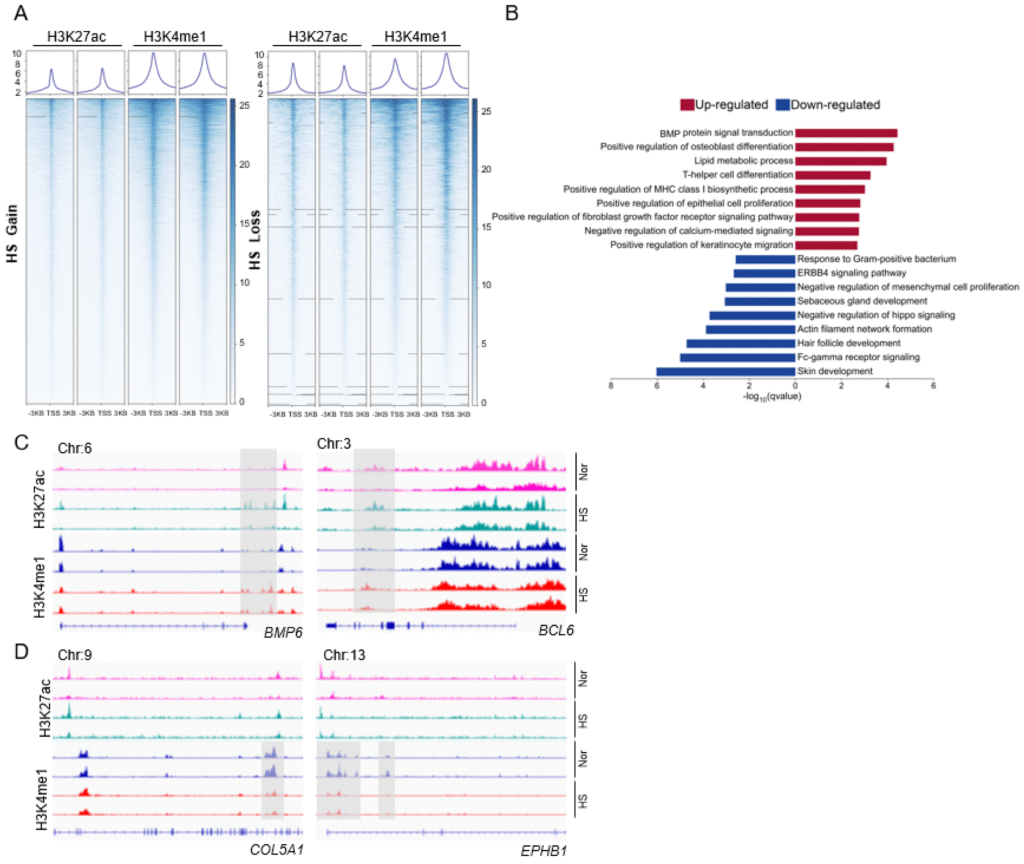
Figure 1: The Characterization of the Epigenetic Regulation Between Healthy and HS Conditions.
- The Scientific Breakdown: Figure by Dr. Lin Jin and colleagues [1]. This figure compares the epigenetic landscape (specifically histone marks H3K27ac and H3K4me1) around gene start sites (TSS) in basal skin cells from healthy individuals versus those with HS.
- Panel A: These heatmaps visually show the gain or loss of these active marks in HS skin. Blue indicates the presence of the mark. You can see distinct patterns where HS skin either gains these marks at certain genes (top) or loses them (bottom) compared to healthy skin.
- Panel B: This chart shows the biological processes (Gene Ontology or GO terms) associated with the genes that gained (red bars) or lost (blue bars) these active epigenetic marks in HS. Genes related to inflammation, cell growth, and specific signaling pathways (like BMP signaling) were turned UP in HS, while genes important for normal skin and hair follicle development were turned DOWN.
- Panels C & D: These are specific examples. They show the epigenetic marks around individual genes like BMP6 (involved in BMP signaling, panel C left), BCL6 (immune cell function, panel C right), COL5A1 (skin structure, panel D left), and EPHB1 (cell signaling, panel D right). The gray boxes highlight regions where the active marks (peaks) are clearly different between normal (Nor) and HS skin.
- What This Means For You: This is like looking at the software code running your skin cells and finding major glitches in HS. The “dimmer switches” are set incorrectly. The instructions for normal skin and hair follicle development seem to be turned down or off (blue bars in B, examples in D), while the instructions for inflammation, abnormal growth, and stress responses are turned way up (red bars in B, examples in C). This isn’t just surface inflammation; the very blueprint for how your skin builds and maintains itself is being epigenetically altered in Hidradenitis Suppurativa causes. It reinforces that HS is a deep cellular problem, not just a plumbing issue with hair follicles.
This epigenetic rewiring suggests that the fundamental instructions for building and maintaining healthy skin are being overridden in HS. Pathways related to normal hair follicle development are silenced, while pathways driving inflammation and abnormal cell behavior (like BMP signaling, which can influence both inflammation and cell growth) are activated. This provides a deep cellular explanation for why HS isn’t just a temporary infection but a chronic condition where the skin itself behaves abnormally.
Reading the Genome: Changes in DNA Accessibility
The researchers also looked at chromatin accessibility using a technique called ATAC-seq. Think of your DNA like a massive library of instruction books. Chromatin is the packaging around these books. Open chromatin means a book is accessible and can be read by the cell’s machinery; “closed chromatin” means it’s packed away.
They found that in HS skin cells, the overall pattern of which DNA books were open and accessible was different compared to healthy cells, particularly in regions containing “transposable elements” (bits of DNA that can sometimes move around) [1].
A Proven natural Roadmap to Manage HS
Get the support and natural strategies you need for lasting relief and join a community that understands.
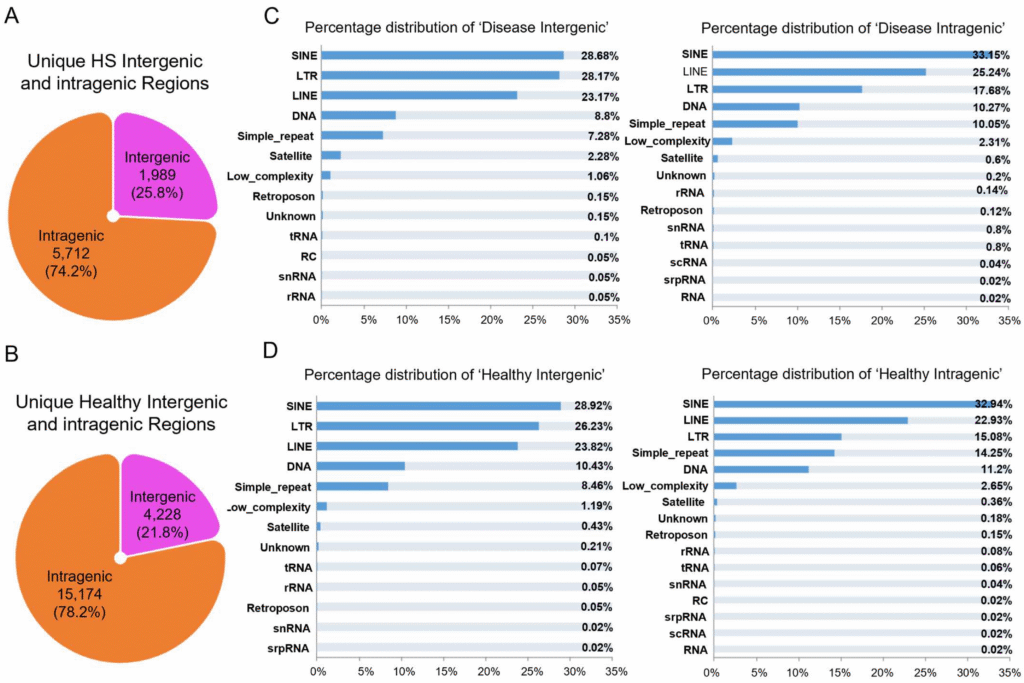
Figure 2: Dynamic Changes in Genome-Wide Chromatin State in HS Condition.
- The Scientific Breakdown: Figure by Dr. Lin Jin and colleagues [1]. This figure examines which parts of the DNA are open or accessible (ATAC-seq peaks) in basal skin cells.
- Panels A & B: These pie charts show the overall distribution of open regions. Interestingly, they found fewer unique open regions in HS skin (A: 1,989 intergenic + 5,712 intragenic = 7,701 total) compared to healthy skin (B: 4,228 intergenic + 15,174 intragenic = 19,402 total), suggesting some parts of the genome might become less accessible in HS. The majority of open regions in both cases are within genes (intragenic).
- Panels C & D: These bar charts break down the types of DNA sequences found within the open regions, focusing on transposable elements (TEs). TEs are repetitive DNA sequences. SINEs, LINEs, and LTRs are major types of retrotransposons (TEs that move via an RNA intermediate). The distribution of these elements within open chromatin was broadly similar between HS (C) and healthy (D) skin, suggesting these elements might influence nearby genes rather than dramatically increasing in number.
- What This Means For You: This adds another layer to the story. Not only are the epigenetic “dimmer switches” different (Figure 1), but the overall accessibility of the DNA “library” also changes in HS. It seems some instruction manuals might be getting packed away or becoming harder to read in HS skin cells. While the details about transposable elements are complex, the main message is that the fundamental way your cells access and read their own DNA instructions is altered in Hidradenitis Suppurativa.
While the exact implications are complex, it adds to the picture of widespread dysregulation at the level of how your cells read and use their own genetic instructions.
A Single-Cell View: Identifying the Troublemakers
To get an even clearer picture, the team used a powerful technique called single-nucleus ATAC-seq (snATAC-seq). This allows them to map the open chromatin library inside individual cells, one by one. It’s like going from a blurry aerial photo of a city to having detailed street maps for every single house.
They identified 13 different clusters, representing various cell types and states within the epidermis (basal cells, spinous cells, hair follicle cells, immune cells like T-cells and Langerhans cells) [1]. Importantly, they found one specific cluster (Cluster 8), representing cells transitioning into the spinous layer, that seemed distinctly altered in HS [1]. This cluster showed open chromatin around genes involved in sending inflammatory signals (cytokines) [1].
A Proven natural Roadmap to Manage HS
Get the support and natural strategies you need for lasting relief and join a community that understands.
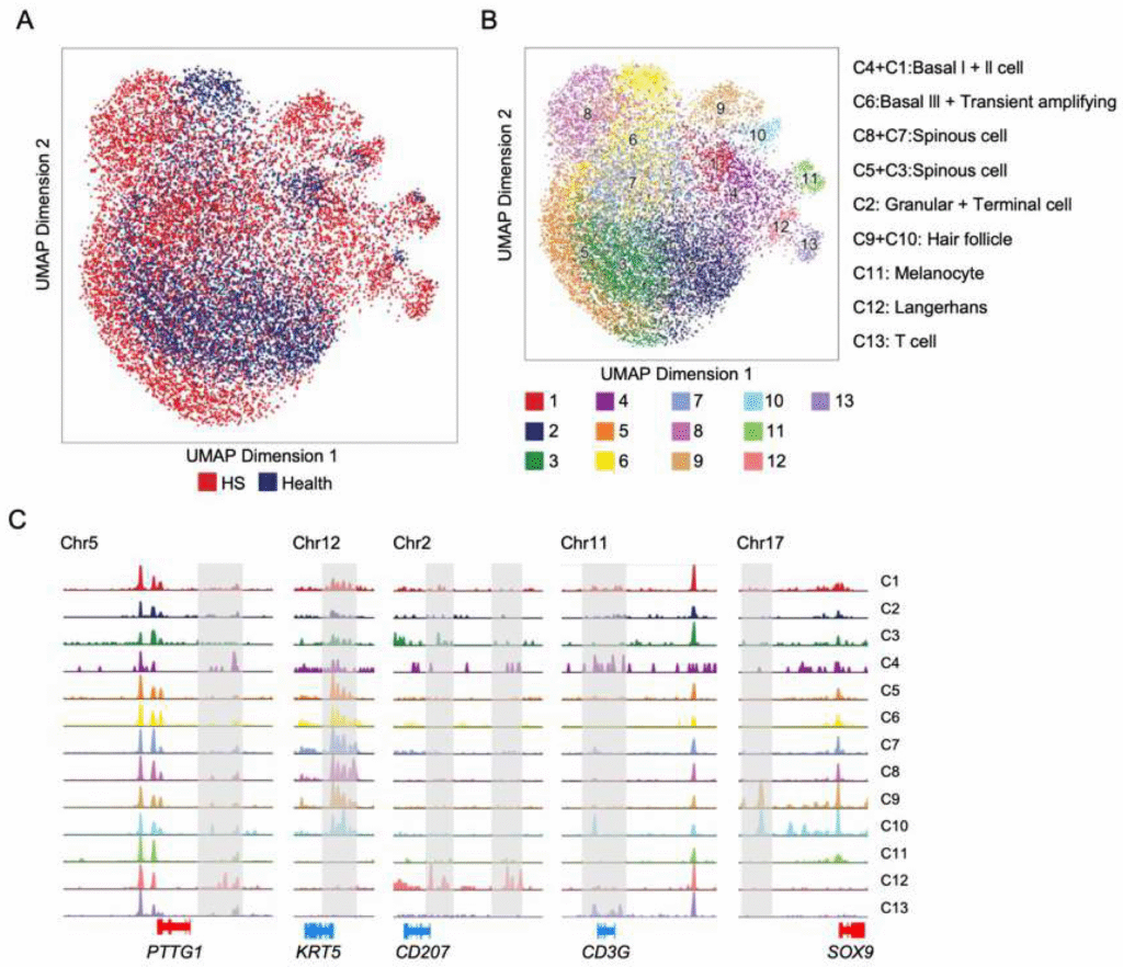
Figure 3: Defining the Chromatin Accessibility of Single Epidermal Nuclei in Healthy and HS Lesional Skin.
- The Scientific Breakdown: Figure by Dr. Lin Jin and colleagues [1]. This figure visualizes the snATAC-seq data, mapping the open chromatin profiles of thousands of individual cells.
- Panel A: This UMAP plot shows all the analyzed cells (nuclei). Each dot is one cell. Red dots are from HS skin, and blue dots are from healthy skin. You can see they mostly overlap, but there are some areas with distinct clustering, suggesting differences in cell states between HS and healthy skin.
- Panel B: This UMAP plot colors the cells based on 13 identified clusters (groups with similar open chromatin patterns). The labels on the right assign potential cell identities to these clusters based on which genes have open chromatin nearby (e.g., Cluster 9+10 are likely Hair Follicle cells because genes like SOX9 are accessible). Cluster 8 is highlighted as potentially HS-specific.
- Panel C: These tracks show the actual snATAC-seq data (peaks representing open DNA) around specific marker genes for different clusters. For example, KRT5 (a keratin gene) has open chromatin in spinous cell clusters (like C7, C8), while CD207 is open in Langerhans cells (C12), and SOX9 in hair follicle cells (C9, C10). This confirms the cell type assignments.
- What This Means For You: This is like creating a high-resolution map of the cellular neighborhoods in your skin. We can see the different types of skin cells and immune cells living there. Crucially, the researchers identified a specific neighborhood (Cluster 8) that looks different and potentially more inflammatory in HS skin compared to healthy skin. This helps pinpoint specific cell states that might be driving the disease process, moving us closer to understanding the cellular origins of Hidradenitis Suppurativa symptoms.
This single-cell view helps us pinpoint specific cell states that might be key drivers of the inflammation in HS.
The ATF3 Connection: A Cellular Stress Signal in Overdrive?
The most intriguing finding came when the researchers looked for specific master switches, transcription factors (TFs), that might be controlling these changes in chromatin accessibility. TFs are proteins that bind to DNA and turn genes on or off.
They focused on two families previously linked to HS inflammation: ATF and NF-κB [1]. Using computational analysis, they predicted the activity of these TFs in each cell cluster.
The results pointed strongly to one TF in particular: ATF3 (Activating Transcription Factor 3) [1].
- ATF3 activity was distinctly increased in the HS-specific cluster (Cluster 8) [1].
- The DNA binding sites (motifs) for ATF3 were highly enriched in the open chromatin regions specific to HS skin, suggesting ATF3 was actively binding and potentially regulating genes in these disease-altered cells [1].
ATF3 is known as a “stress-response” gene. It gets turned on when cells are under pressure – from injury, inflammation, or metabolic stress. Its job is complex and context-dependent; sometimes it helps resolve inflammation, other times it can contribute to it or influence cell growth and remodeling.
A Proven natural Roadmap to Manage HS
Get the support and natural strategies you need for lasting relief and join a community that understands.
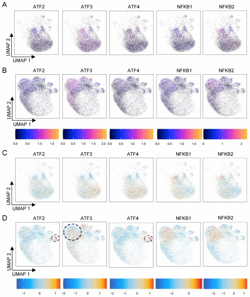
Figure 4: Cell-Type-Specific Transcription Factor (TF) Activity and Its DNA Binding Score.
- The Scientific Breakdown: Figure by Dr. Lin Jin and colleagues [1]. This figure predicts the activity of key inflammation-related transcription factors (ATF family members and NFKB family members) based on the snATAC-seq data.
- Panels A & C (Healthy): These UMAP plots show the predicted TF activity (A) and the predicted TF DNA binding (motif enrichment, C) in healthy skin cells. Darker colors mean higher activity/binding.
- Panels B & D (HS): These plots show the same predictions but for HS skin cells. Compare Panel B to A, and D to C.
- The Key Finding (ATF3): Look specifically at the ATF3 column. In HS skin (B), the activity score for ATF3 is visibly higher, particularly in the upper clusters (including Cluster 8). Even more strikingly, in HS skin (D), the DNA binding score for ATF3 (red/orange indicates higher binding) is strongly enriched in that same upper region (black dotted circle highlights Cluster 8). This suggests ATF3 is not only more active but is specifically binding to open DNA regions in these HS-altered cells. NFKB1/2 also show increased binding in HS (D), consistent with their known role in inflammation. ATF2/4 binding seems increased in the T-cell cluster (gray circle, Cluster 13) in HS (D).
- What This Means For You: Transcription factors are like the managers who decide which instruction manuals (genes) get read. This analysis suggests that in HS skin, a specific manager named ATF3 is working overtime, particularly in the cells identified as potentially problematic (Cluster 8). ATF3 is often activated by cellular stress. This finding provides a molecular link between the feeling of your body being under stress and the actual biological processes happening in your skin cells. It suggests ATF3 might be a key messenger translating systemic stress signals into the cellular misbehavior seen in HS.
The strong signal for ATF3 in HS skin suggests it might be a key player relaying the stress message caused by the underlying inflammation, potentially contributing to the abnormal cell behavior (like hyperproliferation or altered differentiation) that kicks off the HS lesion cycle. NF-κB, a well-known master switch for inflammation, also showed increased activity, confirming the pro-inflammatory state [1].
Seeing is Believing: ATF3 is Increased in HS Skin Tissue
To confirm their computational findings, the researchers performed immunofluorescence staining, using glowing antibodies to visually locate the ATF3 protein in actual skin samples.
The results were clear and confirmed the snATAC-seq predictions [1]:
- ATF3 protein levels were significantly higher throughout the epidermis (outer skin layer) in HS lesions compared to healthy skin [1].
- ATF3 was found not only in the basal layer (where new skin cells are born) but also spread up into the suprabasal layers, co-localizing with KRT14 (a marker for proliferating keratinocytes) [1]. This suggests ATF3 might be involved in the epidermal thickening (hyperplasia) seen in HS.
- ATF3 was also more widespread in the dermis (deeper skin layer) of HS lesions, potentially indicating its role in activated immune cells or fibroblasts (cells involved in scarring) [1].
A Proven natural Roadmap to Manage HS
Get the support and natural strategies you need for lasting relief and join a community that understands.
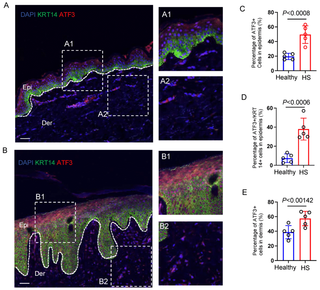
Figure 5: Identification of ATF3 Expression in Healthy and HS Lesional Skin.
- The Scientific Breakdown: Figure by Dr. Lin Jin and colleagues [1]. This figure uses immunofluorescence staining to visually confirm the presence and location of the ATF3 protein in skin tissue.
- Panels A & B: These are microscope images. Green shows KRT14 (marking basal/proliferating skin cells), Red shows ATF3 protein, and Blue shows cell nuclei (DAPI). Panel A is healthy skin; Panel B is HS skin. The white line separates the epidermis (Epi, above) from the dermis (Der, below). The boxes (A1, A2, B1, B2) show magnified views. Notice how much more red staining (ATF3) there is in the HS epidermis (B1) compared to healthy (A1), and how it extends into upper layers. There’s also more ATF3 in the HS dermis (B2 vs. A2).
- Panels C, D, E: These graphs quantify the staining. They confirm significantly higher percentages of ATF3-positive cells in the HS epidermis (C), specifically ATF3 co-localizing with KRT14+ cells (D), and also more ATF3-positive cells in the HS dermis (E), compared to healthy skin.
- What This Means For You: This is the visual proof. The predictions from the fancy computer analysis (Figure 4) hold true in real tissue. The ATF3 protein, that molecular stress signal, is physically present at much higher levels exactly where the HS action is happening – in the thickened epidermis and the inflamed dermis. This strongly supports the idea that ATF3 is not just a bystander but an active participant in the HS disease process, potentially driving abnormal skin cell growth and contributing to the overall inflammation and scarring.
This visual confirmation strongly suggests ATF3 isn’t just a random correlation; it’s likely an active participant in the HS disease process, responding to and perhaps even driving the cellular chaos.
The Knowledge Gap: Silencing the Messenger vs. Stopping the Message
This research is incredibly valuable. Identifying ATF3 as a potential key player opens up exciting possibilities for future drug development. Pharmaceutical companies might look at this and think, “Can we create a drug to block ATF3?”
And that might offer some relief. It would be another tool, another temporary shield. But here lies the critical knowledge gap that defines the difference between conventional treatment and a root-cause approach.
Blocking ATF3 is like trying to silence a messenger who is constantly reporting a fire. It might stop the annoying reports, but it does nothing to extinguish the actual fire. The HS Arma philosophy asks the more fundamental question: Why is ATF3 being activated so strongly? What is the underlying “stress” or “fire” that’s triggering this entire cascade?
The paper itself hints at the answers when discussing the epigenetic changes linked to BMP signaling, T-cell responses, and altered skin development [1]. These are downstream effects. The upstream causes, the true sources of the fire, are the systemic imbalances we focus on:
- Foundational Nutrition: Identifying and removing triggers while providing your body with the nutrient-dense foods it needs to heal and reduce inflammation.
- Strategic Lifestyle Changes: Incorporating practices that reduce systemic inflammation, manage stress, improve sleep, and reduce exposure to environmental toxins and many more.
- Natural Therapies & Skincare: Harnessing the power of nature from both the inside and out. This layer focuses on potent, science-backed natural compounds and supplements that reduce systemic inflammation and support immune balance, key strategies for building your arma internally. This inside-out approach is complemented by targeted natural remedies and skincare routines to soothe the skin, aid wound and scar healing, and manage hidradenitis suppurativa symptoms externally.
- Accountability & Support: Recognizing that this healing journey is challenging. True, lasting change requires personal commitment reinforced by a strong support network. This layer is about staying in contact with Jaap to build resilience, stay on track, and adjust your health plan. Your journey and challenges will also be supported by a community where you can connect with others who understand and can lift you up when you need it most.
- Targeted Medical Testing: We also provide guidance on medical tests you can take to speed up your healing process by identifying key hurdles, triggers, and problems that can arise when dealing with Hidradenitis Suppurativa. We also empower you with information, enabling you to have more effective discussions about treatment options with your medical practitioner.
Conventional medicine often treats these as separate issues or minor contributing factors. We see them as the central, upstream drivers that need to be addressed. We focus on highly effective, evidence-based nutrition and lifestyle changes, and natural therapies and practices. Our goal isn’t just to block a single molecule like ATF3; it’s to create an internal environment so balanced and calm that ATF3 doesn’t need to be activated in the first place. This is how we aim for lasting remission, not just symptom management. Seeing how foundational changes have helped so many in our community achieve remission really brings this science to life.
Key Takeaways
This research is complex, but the core messages for your healing journey are clear:
- HS Involves Deep Cellular Reprogramming: This isn’t just surface inflammation. HS involves changes to your skin cells’ epigenetic software and how they access their DNA library, altering their fundamental behavior.
- ATF3 Emerges as a Key Player: This stress-response transcription factor is significantly upregulated and active in HS skin, potentially acting as a crucial link between systemic stress/inflammation and the abnormal skin cell activity that initiates lesions.
- The “Fire” is Systemic: The activation of ATF3 and the epigenetic changes are likely responses to underlying, body-wide inflammation and stress, confirming HS as a systemic disease.
- Root-Cause Healing is Logical: Targeting downstream molecules like ATF3 might offer temporary relief (a shield), but lasting remission requires addressing the upstream triggers (the fire) through foundational changes in diet, lifestyle, and stress management.
Conclusion: You Hold the Blueprint
This research is incredibly exciting because it takes us deeper than ever before into the cellular mechanics of Hidradenitis Suppurativa. It validates the feeling that this disease is more than skin deep and provides a potential molecular link (ATF3) between the systemic fire and the localized smoke alarm.
So, can you cure HS? While we can’t change our genes, this research powerfully suggests we can change the epigenetic software and the stress signals our cells are receiving. By addressing the root causes of systemic inflammation, the true fire, we can potentially calm down factors like ATF3 and restore a healthier balance to our skin cells.
You are not simply a victim of faulty genes or runaway inflammation. Your body has an incredible capacity for healing. This science doesn’t just offer new drug targets; it offers a deeper understanding that empowers you. By focusing on the foundational pillars of health, nutrition, lifestyle, stress reduction, you are actively working to change the signals your cells receive. You are becoming the architect of your own healing. Remission isn’t just a hope; it’s a biologically plausible outcome when you address the disease at its root.
Reference
- [1] Haque, S., Mohiuddin, S., Khan, J., Muzaffar, S., Vejendla, S., Zhang, Y., Kamata, M., & Jin, L. (2025). Single-Nucleus Chromatin Accessibility and Epigenetic Study Uncover Cell States and Transcriptional Regulation of Epidermis in Hidradenitis Suppurativa. Biomedicines, 13(7), 1599. https://doi.org/10.3390/biomedicines13071599
Important Medical Disclaimer
1. Not Medical Advice: All content and information on this website is for informational and educational purposes only. It does not constitute medical advice and is not a substitute for professional diagnosis, treatment, or consultation with a qualified healthcare provider.
2. My Role and Qualifications: I am a biomedical scientist and PhD candidate and share information from that perspective, combined with my personal experience as a patient with Hidradenitis Suppurativa. However, I am not a medical doctor, physician, or registered healthcare professional. Do not consider our relationship a doctor-patient relationship.
3. Consult Your Doctor: Always seek the advice of your medical doctor or another qualified health professional with any questions you may have regarding a medical condition. Never disregard professional medical advice or delay seeking it because of something you have read on this website. If you suspect you are experiencing a medical emergency, or a severe infection, do not rely on this website or the HS Arma community, please call your local emergency services or go to the nearest emergency room immediately.
4. A Critical Warning on Medication: Pharmaceutical drugs are a crucial tool in managing Hidradenitis Suppurativa for many people. Under absolutely no circumstances should you ever alter, reduce, or stop taking your prescribed medication without the explicit direction of the doctor who prescribed it. Doing so can be dangerous. Always consult with your doctor before doing anything related to your treatment plan.
5. No Liability: Your use of this website and reliance on any information provided is solely at your own risk.
6. Individual Results May Vary: Every patient’s biological baseline, genetics, and adherence to the protocol is different. Therefore, I cannot guarantee specific results, cures, or timelines for your Hidradenitis Suppurativa.
7. Scientific and Expressive Freedom: The articles published on this blog are distinct from formal peer-reviewed academic literature. They serve as an independent platform for my personal viewpoints, scientific hypotheses, and philosophical reflections as an independent scientist and HS patient. While grounded in biomedical research, I exercise a degree of expressive freedom to translate rigid academic data into insights from a patient perspective. These writings are my personal meditations on the science of HS and should be read as my individual perspective, not as universally accepted clinical consensus or formal peer-reviewed literature.